
URL: https://www.desy.de/information__services/press/press_releases/2014/pr_270214/index_eng.html
Breadcrumb Navigation
Researchers X-Ray Living Cancer Cells
Nanodiffraction opens up new insights into the physics of life
")
![Download [0 MB, 1041 x 1043]](/sites2009/site_www-desy/content/e428/e548/e4802/e167391/e167693/e167703/fixed_hydrated_xray_dark_field_ger.jpg)
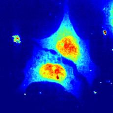
X-ray scan of biological cells: Each pixel represents a complete diffraction image. The colour indicates how strong the X-rays are scattered at this local point. Credit: Britta Weinhausen/University of Göttingen
Hamburg, 27 February 2014. Göttingen-based scientists working at DESY’s PETRA III research light source have carried out the first studies of living biological cells using high-energy X-rays. The new method shows clear differences in the internal cellular structure between living and dead, chemically fixed cells that are often analysed. “The new method for the first time enables us to investigate the internal structures of living cells in their natural environment using hard X-rays,” emphasises the leader of the working group, Prof. Sarah Köster from the Institute for X-Ray Physics of the University of Göttingen. The researchers present their work in the scientific journal Physical Review Letters.
Thanks to analytical methods with ever-higher resolution, scientists today can study biological cells at the level of individual molecules. The cells are frequently chemically fixed before they are studied with the help of optical, X-ray or electron microscopes. The process of chemical fixation involves immersing the cells in a type of chemical preservative which fixes all of the cell’s organelles and even the proteins in place. “Minor changes to the internal structure of the cells are unavoidable in this process,” emphasises Köster. “In our studies, we were able to show these changes in direct comparison for the first time.”
The team used cancer cells from the adrenal cortex for their analyses. They grew the cells on a silicon nitrite substrate, which is almost transparent to X-rays. In order to keep the cells alive in the experimental chamber during the experiment, they were supplied with nutrients, and their metabolic products were pumped away via fine channels just 0.5 millimetres in diameter. “The biological cells are thus located in a sample environment which very closely resembles their natural environment,” explains Dr. Britta Weinhausen from Köster’s group, the paper’s first author.
The experiments were carried out at the Nanofocus Setup (GINIX) of PETRA III’s experimental station P10. The scientists used the brilliant X-ray beam from PETRA III to scan the cells in order to obtain information about their internal nanostructure. “Each frame was exposed for just 0.05 seconds, in order to avoid damaging the living cells too quickly”, explains co-author Dr. Michael Sprung from DESY. “Even nanometre-scale structures can be measured with the GINIX assembly, thanks to the combination of PETRA III’s high brilliance and the GINIX setup which is matched to the source.”
The researchers studied living and chemically fixed cells using this so-called nanodiffraction technique and compared the cells’ internal structures on the basis of the X-ray diffraction images. The results showed that the chemical fixation produces noticeable differences in the cellular structure on a scale of 30 to 50 nanometres (millionths of a millimetre).
“Thanks to the ever-greater resolution of the various investigative techniques, it is increasingly important to know whether the internal structure of the sample changes during sample preparation,” explains Köster. In future, the new technique will make it possible to study unchanged living cells at high resolution. Although other methods have an even higher resolution than X-ray scattering, they require a chemical fixation or complex and invasive preparation of the cells. Lower-energy, so-called soft X-rays have already been used for studies of living cells. However, the study of structures with sizes as small as 12 nanometres first becomes possible through the analysis of diffraction images produced using hard X-rays.
Deutsches Elektronen-Synchrotron DESY is the leading German accelerator centre and one of the leading in the world. DESY is a member of the Helmholtz Association and receives its funding from the German Federal Ministry of Education and Research (BMBF) (90 percent) and the German federal states of Hamburg and Brandenburg (10 percent). At its locations in Hamburg and Zeuthen near Berlin, DESY develops, builds and operates large particle accelerators, and uses them to investigate the structure of matter. DESY’s combination of photon science and particle physics is unique in Europe.
Reference
“Scanning X-ray Nano-Diffraction on Living Eukaryotic Cells in Microfluidic Environments”; Britta Weinhausen et al.; Physical Review Letters, 2014; DOI: 10.1103/PhysRevLett.112.088102
Science contacts
Prof. Sarah Köster, University of Göttingen, +49 551 39-5556, Sarah.Koester@phys.uni-goettingen.de
Dr. Michael Sprung, DESY, +49 40 8998-4680, michael.sprung@desy.de
Media contact
DESY press officer Thomas Zoufal, +49 8998-1666, presse@desy.de
Images
")
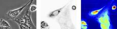
Visible light phase contrast microscopy image (left), fluorescence microscopy image (middle) and X-ray scan (right) of chemically fixed cells. Credit: Britta Weinhausen/University of Göttingen |
")
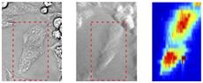
Visible light phase contrast microscopy image (left), fluorescence microscopy image (middle) and X-ray scan (right) of living cells. To avoid damaging the cells too soon, the scan resolution was reduced. However, the spatial resolution of the structural information retrieved from the diffraction pattern remains the same. Credit: Britta Weinhausen/University of Göttingen
|
")
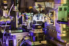
The GINIX set-up at beamline P10 at PETRA III. Just right of the picture's centre the test chamber can be seen. Credit: Markus Osterhoff/University of Göttingen |
![Download [0 MB, 3198 x 811]](/sites2009/site_www-desy/content/e428/e548/e4802/e167391/e167693/e167695/column-objekt167697/img/bw145_ger.jpg)
![Download [0 MB, 2921 x 1203]](/sites2009/site_www-desy/content/e428/e548/e4802/e167391/e167693/e167695/column-objekt167698/img/living_cell_ger.jpg)
![Download [2 MB, 4252 x 2835]](/sites2009/site_www-desy/content/e428/e548/e4802/e167391/e167693/e167695/column-objekt168773/img/EH7Q8021_ger.jpg)