
URL: https://www.desy.de/news/news_search/index_eng.html
Breadcrumb Navigation
DESY News: Table-top electron camera catches ultrafast dynamics of matter
News
News from the DESY research centre
Table-top electron camera catches ultrafast dynamics of matter
Scientists at DESY have built a compact electron camera that can capture the inner, ultrafast dynamics of matter. The system shoots short bunches of electrons at a sample to take snapshots of its current inner structure and is the first such electron diffractometer that uses Terahertz radiation for pulse compression. The developer team around DESY scientists Dongfang Zhang and Franz Kärtner from the Center for Free-Electron Laser Science CFEL validated their Terahertz-enhanced ultrafast electron diffractometer with the investigation of a silicon sample and present their work in the first issue of the journal Ultrafast Science, a new title in the Science group of scientific journals.
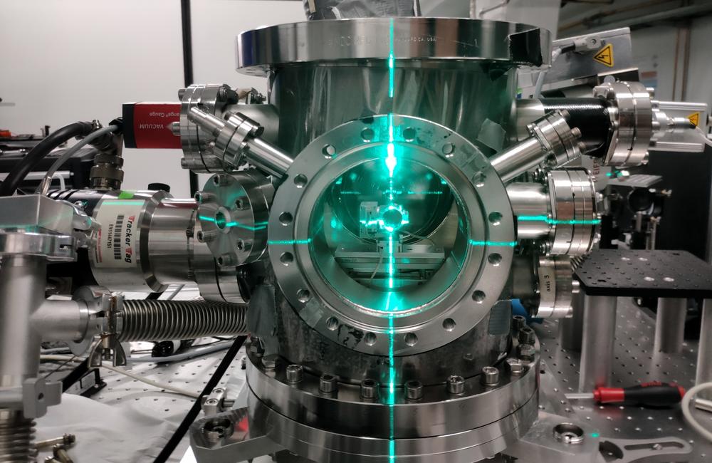
The system fits on a lab table. It is adjusted with the help of an optical laser (green). Credit: DESY, Timm Rohwer
Such short electron bunches can be routinely produced with high quality by state-of-the-art particle accelerators. However, these machines are often large and bulky, partly due to the radio frequency radiation used to power them, which operates in the Gigahertz band. The wavelength of the radiation sets the size for the whole device. The DESY team is now using Terahertz radiation instead with roughly a hundred times shorter wavelengths. “This basically means, the accelerator components, here a bunch compressor, can be a hundred times smaller, too,” explains Kärtner, who is also a professor and a member of the cluster of excellence “CUI: Advanced Imaging of Matter“ at the University of Hamburg.
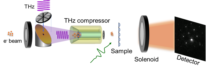
On top of its reduced size, the Terahertz electron diffractometer has another advantage that might be even more important to researchers: “Our system is perfectly synchronised, since we are using just one laser for all steps: generating, manipulating, measuring and compressing the electron bunches, producing the Terahertz radiation and even heating the sample,” Kärtner explains. Synchronisation is key in this kind of ultrafast experiments. To monitor the swift structural changes within a sample of matter like silicon, researchers usually repeat the experiment many times while delaying the measuring pulse a little more each time. The more accurate this delay can be adjusted, the better the result. Usually, there needs to be some kind of synchronisation between the exciting laser pulse that starts the experiment and the measuring pulse, in this case the electron bunch. If both, the start of the experiment and the electron bunch and its manipulation are triggered by the same laser, the synchronisation is intrinsically given.
In a next step, the scientists plan to increase the energy of the electrons. Higher energy means the electrons can penetrate thicker samples. The prototype set-up used rather low-energy electrons and the silicon sample had to be sliced down to a thickness of just 35 nanometres (millionths of a millimetre). Adding another acceleration stage could give the electrons enough energy to penetrate 30 times thicker samples with a thickness of up to 1 micrometre (thousandth of a millimetre), as the researchers explain. For even thicker samples, X-rays are normally used. While X-ray diffraction is a well established and hugely successful technique, electrons usually do not damage the sample as quickly as X-rays do. “The energy deposited is much lower when using electrons,“ explains Zhang. This could prove useful when investigating delicate materials.
This work has been supported by the European Research Council under the European Union’s Seventh Framework Program (FP7/2007-2013) through the Synergy Grant AXSIS (609920), Project KA908-12/1 of the Deutsche Forschungsgemeinschaft, and the accelerator on a chip program (ACHIP) funded by the Gordon and Betty Moore foundation (GBMF4744).
Reference
THz-Enhanced DC Ultrafast Electron Diffractometer; Dongfang Zhang, Tobias Kroh, Felix Ritzkowsky, Timm Rohwer, Moein Fakhari, Huseyin Cankaya, Anne-Laure Calendron, Nicholas H. Matlis, and Franz X. Kärtner; Ultrafast Science, 2021; DOI: 10.34133/2021/9848526
