
URL: https://www.desy.de/news/news_search/index_eng.html
Breadcrumb Navigation
DESY News: Roentgen Medal 2017 for Henry Chapman
News
News from the DESY research centre
Roentgen Medal 2017 for Henry Chapman
DESY scientist Henry Chapman has been awarded the Roentgen Medal by the city of Remscheid. The town in which Wilhelm Conrad Roentgen was born has been presenting this award annually since 1951 to individuals who have made outstanding contributions towards improving and advancing the use of the radiation discovered by Roentgen. Henry Chapman, a Leading Scientist at DESY and professor at the University of Hamburg, has been awarded the Medal in recognition of his pioneering work on the application of X-ray lasers for determining the structure of biological macromolecules.
Henry Chapman is head of the department for Coherent X-Ray Imaging at the Center for Free-Electron Laser Science, jointly operated by DESY, the University of Hamburg and the Max Planck Society. Chapman’s team develops sophisticated new experimental techniques for making use of the excellent new opportunities offered by free-electron lasers (FEL). The X-ray pulses produced by these devices can be up to a billion times more intense and a thousand times shorter than those from conventional synchrotron radiation sources.
The aim is to use the laser-like beams to see the structure and the dynamics of proteins and their complexes—the molecular machines of life. But doing so calls for entirely new equipment and methods of 3D imaging, such as X-ray holography and atomic-scale diffraction tomography.
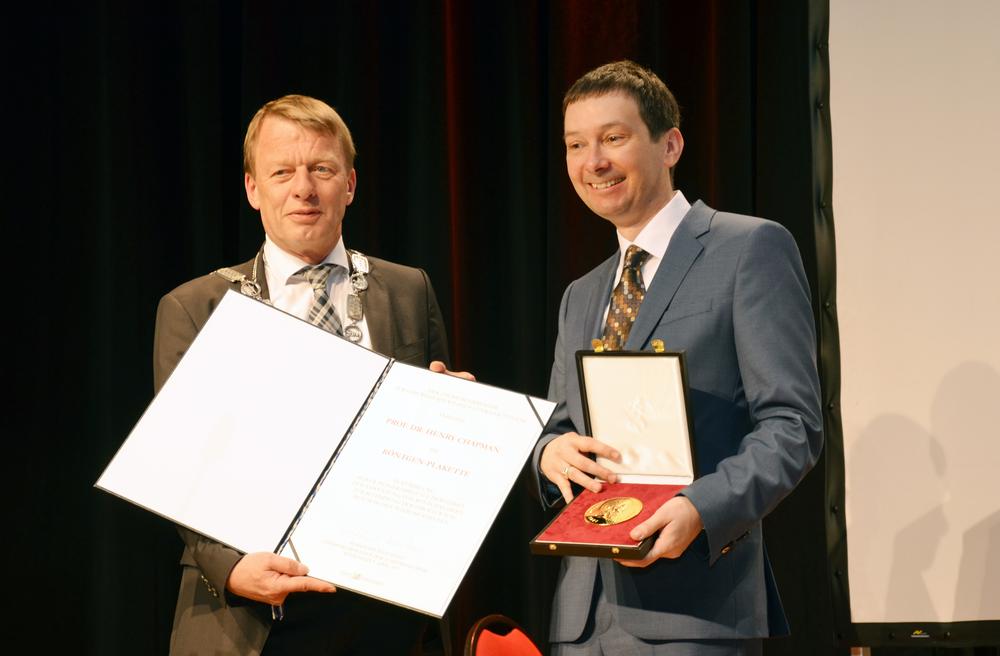
Remscheid's Mayor Burkhard Mast-Weisz (left) presented Chapman with the medal. Credit: Deutsches Röntgen-Museum, Remscheid
This method called “diffraction before destruction” overcomes previous limitations to studying such materials with X-ray beams, and forms the basis for exploiting the full research potential of the new generation of X-ray lasers, such as the European XFEL, which is to go into operation this year. Chapman has already used the method, for example, to establish the structure of a parasitic protein, the enzyme cathepsin B, in the pathogen that causes tropical sleeping sickness.
Another breakthrough achieved by Chapman was in the field of conventional X-ray crystallography: in 2016, he and his team discovered a method for deciphering the structure of complex biomolecules at high resolutions, without the need for the perfectly ordered crystals hitherto required. Contrary to the established wisdom, he showed that slightly disordered crystals, which are much easier to grow and prepare, actually give superior information for unravelling the structure of proteins.
